Overview
Although tibial
plateau fracture was originally termed a bumper or fender fracture, only
25% of tibial plateau fractures result from impact with automobile
bumpers. The most common mechanism of injury involves axial loading,
such as results from a fall. Other patterns of injury result from
laterally directed forces or from a twisting injury. In all cases, force
is directed from the femoral condyles onto the medial and lateral
portions of the tibial plateau, resulting in fracture. In younger
patients, the most common pattern of fracture is splitting, while in
older, more osteoporotic patients, depression fractures typically are
sustained.
Examples of tibial plateau fractures are provided in the images below:
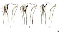 Tibial
plateau fractures. Line drawings of Schatzker types I, II, and III
tibial plateau fractures. Type I consists of a wedge fracture of the
lateral tibial plateau, produced by low-force injuries. Type II combines
the wedge fracture of the lateral plateau with depression of the
lateral plateau. Type III fractures are classified as those with
depression of the lateral plateau but no associated wedge fracture.
Tibial
plateau fractures. Line drawings of Schatzker types I, II, and III
tibial plateau fractures. Type I consists of a wedge fracture of the
lateral tibial plateau, produced by low-force injuries. Type II combines
the wedge fracture of the lateral plateau with depression of the
lateral plateau. Type III fractures are classified as those with
depression of the lateral plateau but no associated wedge fracture. 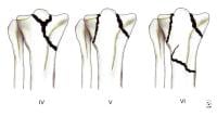 Tibial
plateau fractures. Line drawings of Schatzker types IV, V, and VI
tibial plateau fractures. Type IV is similar to type I fracture, except
that it involves the medial tibial plateau as opposed to the lateral
plateau. Greater force is required to produce this type of injury. Type V
fractures are termed bicondylar and demonstrate wedge fractures of both
the medial and lateral tibial plateaus. Finally, type VI fractures
consist of a type V fracture along with a fracture of the underlying
diaphysis and/or metaphysis.
Tibial
plateau fractures. Line drawings of Schatzker types IV, V, and VI
tibial plateau fractures. Type IV is similar to type I fracture, except
that it involves the medial tibial plateau as opposed to the lateral
plateau. Greater force is required to produce this type of injury. Type V
fractures are termed bicondylar and demonstrate wedge fractures of both
the medial and lateral tibial plateaus. Finally, type VI fractures
consist of a type V fracture along with a fracture of the underlying
diaphysis and/or metaphysis. 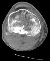 Tibial
plateau fractures. CT image through the tibial plateau shows a fracture
of the posterior aspect of the lateral tibial plateau, which is the
source of the lipohemarthrosis.
Tibial
plateau fractures. CT image through the tibial plateau shows a fracture
of the posterior aspect of the lateral tibial plateau, which is the
source of the lipohemarthrosis. 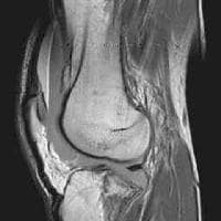 Tibial
plateau fractures. MRI of the knee in a patient with tibial plateau
fracture and lipohemarthrosis. Three layers of effusion are demonstrated
on this proton density sequence: fat, red blood cells, and serum.
Low-signal intensity in the tibial plateau corresponds to the site of
fracture.
Tibial
plateau fractures. MRI of the knee in a patient with tibial plateau
fracture and lipohemarthrosis. Three layers of effusion are demonstrated
on this proton density sequence: fat, red blood cells, and serum.
Low-signal intensity in the tibial plateau corresponds to the site of
fracture. 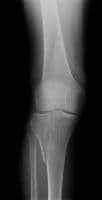 Tibial
plateau fractures. Radiograph of the knee shows lateral plateau
splitting, a Schatzker I injury. There is no articular depression. Soft
tissue injuries (eg, to cruciate and collateral ligaments) occur in
approximately 10% of patients. In particular, medial plateau injuries
may result in fracture of the fibular head, which may injure the
peroneal nerve or may be associated with popliteal artery occlusion.
Patients may present with a knee effusion, pain, and joint stiffness.
Finally, although severe fractures often are repaired surgically, both
operatively and nonoperatively treated fractures are at risk of
developing posttraumatic osteoarthritis as a result of ligamentous
injuries with resultant instability as well as articular discongruities,
biomechanical alteration of normal compressive forces, and cartilage
damage.
Tibial
plateau fractures. Radiograph of the knee shows lateral plateau
splitting, a Schatzker I injury. There is no articular depression. Soft
tissue injuries (eg, to cruciate and collateral ligaments) occur in
approximately 10% of patients. In particular, medial plateau injuries
may result in fracture of the fibular head, which may injure the
peroneal nerve or may be associated with popliteal artery occlusion.
Patients may present with a knee effusion, pain, and joint stiffness.
Finally, although severe fractures often are repaired surgically, both
operatively and nonoperatively treated fractures are at risk of
developing posttraumatic osteoarthritis as a result of ligamentous
injuries with resultant instability as well as articular discongruities,
biomechanical alteration of normal compressive forces, and cartilage
damage.
 Tibial
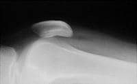
plateau fractures. Cross-table lateral radiograph of the knee shows the
lipohemarthrosis within the suprapatellar bursa. The fracture itself is
not seen well. Tibial
plateau fractures. CT image through the tibial plateau shows a fracture
of the posterior aspect of the lateral tibial plateau, which is the
source of the lipohemarthrosis.
Tibial
plateau fractures. Cross-table lateral radiograph of the knee shows the
lipohemarthrosis within the suprapatellar bursa. The fracture itself is
not seen well. Tibial
plateau fractures. CT image through the tibial plateau shows a fracture
of the posterior aspect of the lateral tibial plateau, which is the
source of the lipohemarthrosis. 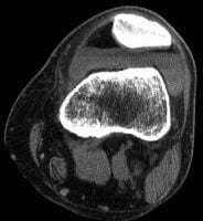 Tibial plateau fractures. Axial CT image through the knee shows a layering lipohemarthrosis. Tibial
plateau fractures. MRI of the knee in a patient with tibial plateau
fracture and lipohemarthrosis. Three layers of effusion are demonstrated
on this proton density sequence: fat, red blood cells, and serum.
Low-signal intensity in the tibial plateau corresponds to the site of
fracture. CT is used by most orthopedists to further
characterize fractures of the tibial plateau and assess the depression
of the tibia and the degree of diastasis (splitting) of the fractured
parts to plan for surgical intervention. Generally, slice thickness
should be minimized (1 mm is ideal) and high milliamperage-second (mAs)
technique used.[1, 2, 3]
Tibial plateau fractures. Axial CT image through the knee shows a layering lipohemarthrosis. Tibial
plateau fractures. MRI of the knee in a patient with tibial plateau
fracture and lipohemarthrosis. Three layers of effusion are demonstrated
on this proton density sequence: fat, red blood cells, and serum.
Low-signal intensity in the tibial plateau corresponds to the site of
fracture. CT is used by most orthopedists to further
characterize fractures of the tibial plateau and assess the depression
of the tibia and the degree of diastasis (splitting) of the fractured
parts to plan for surgical intervention. Generally, slice thickness
should be minimized (1 mm is ideal) and high milliamperage-second (mAs)
technique used.[1, 2, 3]
MRI may be used as well for this determination but often is not readily available. MRI is excellent for depicting ligamentous and meniscal injuries.
Arteriography (and possibly MR angiography) may be used if popliteal artery injury is suspected.[4]
According to Mustonen et al, although postoperative multidetector-row CT (MDCT) scanning of tibial plateau fractures is performed infrequently, it can in most cases reveal clinically significant information. In their study, the main indications for MDCT were assessment and follow-up of the joint articular surface and evaluation of fracture healing. Postoperative MDCT revealed additional clinically important information in 81% of patients, and 39% underwent reoperation. Orthopedic hardware caused no diagnostic problems with MDCT.[6]
Examples of tibial plateau fractures are provided in the images below:
Tibial
plateau fractures. Line drawings of Schatzker types I, II, and III
tibial plateau fractures. Type I consists of a wedge fracture of the
lateral tibial plateau, produced by low-force injuries. Type II combines
the wedge fracture of the lateral plateau with depression of the
lateral plateau. Type III fractures are classified as those with
depression of the lateral plateau but no associated wedge fracture. Tibial
plateau fractures. Line drawings of Schatzker types IV, V, and VI
tibial plateau fractures. Type IV is similar to type I fracture, except
that it involves the medial tibial plateau as opposed to the lateral
plateau. Greater force is required to produce this type of injury. Type V
fractures are termed bicondylar and demonstrate wedge fractures of both
the medial and lateral tibial plateaus. Finally, type VI fractures
consist of a type V fracture along with a fracture of the underlying
diaphysis and/or metaphysis. Tibial
plateau fractures. CT image through the tibial plateau shows a fracture
of the posterior aspect of the lateral tibial plateau, which is the
source of the lipohemarthrosis. Tibial
plateau fractures. MRI of the knee in a patient with tibial plateau
fracture and lipohemarthrosis. Three layers of effusion are demonstrated
on this proton density sequence: fat, red blood cells, and serum.
Low-signal intensity in the tibial plateau corresponds to the site of
fracture. Tibial
plateau fractures. Radiograph of the knee shows lateral plateau
splitting, a Schatzker I injury. There is no articular depression. Soft
tissue injuries (eg, to cruciate and collateral ligaments) occur in
approximately 10% of patients. In particular, medial plateau injuries
may result in fracture of the fibular head, which may injure the
peroneal nerve or may be associated with popliteal artery occlusion.
Patients may present with a knee effusion, pain, and joint stiffness.
Finally, although severe fractures often are repaired surgically, both
operatively and nonoperatively treated fractures are at risk of
developing posttraumatic osteoarthritis as a result of ligamentous
injuries with resultant instability as well as articular discongruities,
biomechanical alteration of normal compressive forces, and cartilage
damage.Preferred examination
The preferred examination consists of radiographs in multiple obliquities of the knee. Typically, these include anteroposterior (AP), cross-table lateral, patellar (sunrise), and, possibly, oblique views. Cross-table lateral and AP may be the only views possible in the trauma suite. In this setting, the cross-table lateral radiograph may be the most important to detect occult fractures. The presence of these subtle fractures may be inferred by the presence of a lipohemarthrosis on the cross-table lateral radiograph, indicating disruption of an articular surface, most often the tibia. The images below demonstrate the radiographic, computed tomography (CT), and magnetic resonance imaging (MRI) appearance of lipohemarthrosis.Tibial
plateau fractures. Cross-table lateral radiograph of the knee shows the
lipohemarthrosis within the suprapatellar bursa. The fracture itself is
not seen well. Tibial
plateau fractures. CT image through the tibial plateau shows a fracture
of the posterior aspect of the lateral tibial plateau, which is the
source of the lipohemarthrosis. Tibial plateau fractures. Axial CT image through the knee shows a layering lipohemarthrosis. Tibial
plateau fractures. MRI of the knee in a patient with tibial plateau
fracture and lipohemarthrosis. Three layers of effusion are demonstrated
on this proton density sequence: fat, red blood cells, and serum.
Low-signal intensity in the tibial plateau corresponds to the site of
fracture. CT is used by most orthopedists to further
characterize fractures of the tibial plateau and assess the depression
of the tibia and the degree of diastasis (splitting) of the fractured
parts to plan for surgical intervention. Generally, slice thickness
should be minimized (1 mm is ideal) and high milliamperage-second (mAs)
technique used.[1, 2, 3] MRI may be used as well for this determination but often is not readily available. MRI is excellent for depicting ligamentous and meniscal injuries.
Arteriography (and possibly MR angiography) may be used if popliteal artery injury is suspected.[4]
Limitations of techniques
Nondepressed tibial plateau fractures occasionally are difficult to appreciate with standard radiographs. Cross-table lateral radiographs may demonstrate a lipohemarthrosis within the joint, with layering of bone marrow fat upon blood. If lipohemarthrosis is present, an intra-articular fracture is present and must be located. In this situation, axial CT is an excellent tool for defining fracture anatomy using reconstructed images in the sagittal and coronal planes.Recent studies
Brunner et al found that CT scanning improved the interobserver and intraobserver reliability of the Schatzker, OTA/AO, and Hohl classification systems for tibial plateau fractures. The 3 systems showed moderate interobserver reliability and good and moderate intraobserver reliability when based only on findings on plain radiographs. Interobserver and intraobserver reliability improved significantly when CT was added.[5]According to Mustonen et al, although postoperative multidetector-row CT (MDCT) scanning of tibial plateau fractures is performed infrequently, it can in most cases reveal clinically significant information. In their study, the main indications for MDCT were assessment and follow-up of the joint articular surface and evaluation of fracture healing. Postoperative MDCT revealed additional clinically important information in 81% of patients, and 39% underwent reoperation. Orthopedic hardware caused no diagnostic problems with MDCT.[6]
No comments:
Post a Comment